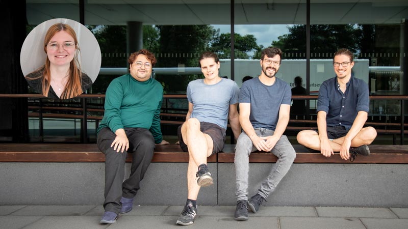
Five of eleven nominated DIGS-ILS PhD students received the DIGS-ILS Fellow Award 2025:
- Simon Doll (Marcus Jahnel Group)
- Waqar Hussain (Claudia Ball Group)*
- Adéla Karhanová (Zdeněk Lánský Group, CAS Prague – as part of the 1st TPC Cytoskeletal Dynamics Across Scales)*
- Lennart Kleinschmidt (Alf Honigmann Group)
- Lukas Niese (Stefan Diez Group)
The award honours outstanding PhD students after the 1st year of thesis work and includes a price money of €2,000 (* split prize this year). It aims to help their research work, development of research skills, and to strengthen their research network. The allowance can be used to cover project consumables, attendance to workshops and conferences, research visits to collaborators’ labs, as well as to cover cost for a side project.
An outstanding performance during the 1st year of PhD work, along with a nomination and justification by their Thesis Advisory Committee (TAC) in the 1st AR TAC meeting is required to be eligible for the award. The students presented the current state of their thesis work to the DIGS-ILS Steering Committee in writing. Applications positively evaluated were invited to defend their application in a chalk talk before the committee. Detailed information on that work can be found below.
Simon Doll
Capillary forces between biomolecular condensates and biopolymers
In my project, I try to understand how condensates of RNA binding proteins can change the folding dynamics and structure of long non-coding RNA. I am especially interested in the question, what role phase separation played during the evolution of the ribosome. Being a fundamental building block of all living cells, parts of its structure and interactions with ribosomal proteins are assumed to be older than the last universal common ancestor - over 3.5 billion years.To tackle this challenge, I am applying bioinformatics, theoretical biophysics and single molecule experiments. I hope, that studying the influence of condensates on the dynamics of the ribosomal structure opens a unique window into the origin of what we call life.
Waqar Hussain
Exploring Molecular Landscapes and Heterogeneity in Rare Cancers
A key challenge in treating cancers lies in heterogeneity; the idea that tumors often exhibit distinct molecular characteristics, not only among different individuals but also between different tumors of the same individual; and even within a single tumor, genetically distinct subpopulations can co-exist. This explains that the heterogeneity exists at various levels, such as inter-patient, inter-tumour, and intra-tumour, each with distinct biological and clinical implications. Inter-patient heterogeneity refers to differences in tumor biology among individuals, influenced by factors such as genetic background, whereas the inter-tumour heterogeneity could be described as variation between multiple tumors (i.e. primary and metastasis) within the same individual, differing mainly in genetic alterations, molecular profiles and therapeutic responses. At a deeper level, intra-tumour heterogeneity (ITH), a major contributor to therapy resistance, is characterised by the co-existence of multiple subclones within a single tumor. These subclonal populations of cancer cells evolve through various genetic and epigenetic alterations, complicating treatment strategies, accelerating the disease progression and its recurrence. Studying heterogeneity gets more complex when the cancer itself is rare, due to less established research and resources. Rare cancers, with occurrence of <6/100,000 cases per year, form a fragmented group of malignancies, responsible for 22% of all human cancers, yet 30% of all cancer-related fatalities. My project investigates how molecular landscapes of these rare cancers contribute to tumor heterogeneity. I integrate comprehensive cohort analyses of multiomics bulk sequencing data such as genomics/epigenomics, transcriptomics, and proteomics, which reveal inter-patient/inter-tumour heterogeneity, with granular details of intra-tumour heterogeneity assessed through single-cell DNA sequencing (scDNA-seq). This high-resolution approach allows me to map clonal architecture and evolutionary trajectories at unprecedented depth, revealing how tumors evolve over time and to study the dynamics of therapy resistance development. With the help of state-of-the-art technologies such as scDNA-seq and integrative multiomics, I characterise the heterogeneity in order to find important genetic causes, find new biomarkers, and shed light on the evolution of tumors. My project improves patient classification, improving precision medicine, and eventually could lead to more individualised and successful treatment plans for these difficult-to-treat, rare cancers.
Adéla Karhanová
Regulation of tau envelopes by post-translational modifications
Microtubules are a cytoskeletal component involved in many cellular functions—such as cell division, maintenance of cell shape, cell motility, and intracellular transport. To perform this wide variety of tasks, microtubules are regulated by associated proteins. These proteins can alter microtubule dynamics and, through competition and mutual interactions, shape cellular processes. For example, unstructured microtubule-associated proteins—such as tau—can form cohesive envelopes around microtubules. These envelopes selectively modulate microtubule accessibility by locally excluding certain proteins from the microtubule surface (e.g., preventing katanin from severing the microtubule) while recruiting others. Posttranslational modifications of tau direct its function in physiological contexts (e.g., during mitosis), and dysregulation of these modifications can lead to diseases such as Alzheimer’s disease or frontotemporal dementia. The aim of my PhD project is to elucidate the mechanisms that modulate tau–microtubule interactions, as these are essential for tau’s roles in both physiology and disease.
Lennart Kleinschmidt
Investigating pattern formation of epithelial adhesion receptors by in vitro reconstitution
In my project, I aim to test the hypothesis that size segregation of adhesion receptors contributes to the organization and function of the apical junctional complex, a key structure of epithelial tissues that regulates cell polarity, mediates cell-cell adhesion and seals the tissue by restricting paracellular diffusion. Therefore, I use a bottom-up in vitro reconstitution of cell-cell interfaces using adhesion of giant unilamellar vesicles (GUVs) on supported lipid bilayers (SLBs). These membranes can be coated with purified receptor ecto-domains (JAM-A, Nectin-1, E-Cadherin), enabling us to quantify protein patterning at the adhesion interface via fluorescence microscopy. With this approach, we are able to assess ecto-domain driven receptor organization and to test the effect of size differences by truncating these domains. Based on our results, we aim establish a biophysical model that predicts the size-dependent patterning of adhesion receptors in epithelial tissues.
Lukas Niese
Torque generation by molecular motors
Multiple motor proteins, including dynein and kinesins 2, 4, 5 and 14, have been observed not only to move straight along microtubules, but also to feature an additional sideways component, leading to helical pathways around a microtubule. This may be a means of avoiding obstacles on individual microtubules and a way of generating lateral force and torque at microtubule overlaps. It has been proposed that motor sidestepping introduces torque in the mitotic spindle, achieving symmetry breaking and creating a more robust chiral structure. I seek to unravel the molecular mechanism behind motor sidestepping and the creation of torsional forces in microtubule overlaps, and to quantify the force potential of such systems in vitro.
